How to Read an EKG Step by Step
Learn the basics of EKG interpretation using a simple step-by-step method. This beginner-friendly guide covers heart rate, rhythm, waveforms, and common rhythm patterns for study and exam preparation.
Start with pattern recognition first. Learn what normal looks like, then compare each rhythm by rate, regularity, P waves, and QRS width.
Important Note
This guide is for educational purposes only. It is designed to help students and learners understand EKG basics and pattern recognition. It does not provide medical certification, clinical training, diagnosis, treatment advice, or professional credentialing.
What is an EKG?
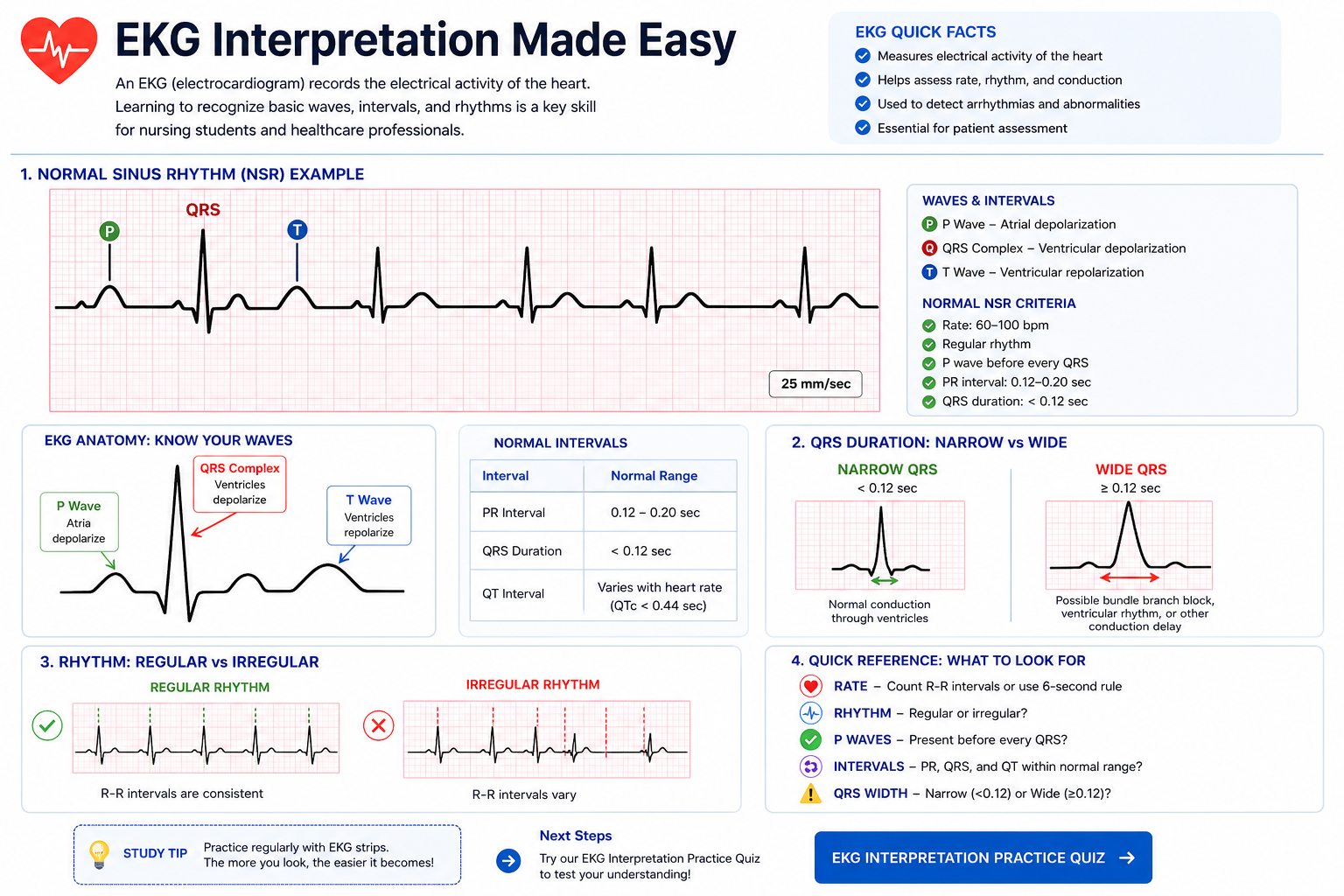
An electrocardiogram, commonly called an EKG or ECG, records the electrical activity of the heart. It helps show how the heart is beating, whether the rhythm is regular or irregular, and whether certain patterns appear normal or abnormal.
For beginners, the key is not to memorize everything at once. The best approach is to follow the same process every time you look at a rhythm strip.
Basic EKG Components
- P Wave: Represents atrial depolarization, or atrial electrical activity before contraction.
- QRS Complex: Represents ventricular depolarization, which leads to ventricular contraction.
- T Wave: Represents ventricular repolarization, or recovery.
- PR Interval: Helps show conduction time from the atria to the ventricles.
- QRS Width: Helps you decide whether conduction is narrow and likely supraventricular or wide and more likely ventricular.
Do not start by trying to name every rhythm. Start by asking: Is it fast or slow? Regular or irregular? Are P waves present? Is the QRS narrow or wide?
Simple Step by Step Approach
1. Check the heart rate
Start by deciding whether the rate is slow, normal, or fast. This quickly narrows the possibilities and helps separate bradycardia, normal sinus rhythm, and tachycardia patterns.
2. Assess rhythm regularity
Look at the spacing between beats. If the spacing is even, the rhythm is regular. If the spacing changes, the rhythm is irregular.
3. Look for P waves
Check whether a P wave appears before each QRS complex. Clear P waves often suggest the rhythm is following the normal sinus pathway.
4. Evaluate the QRS complex
Decide whether the QRS is narrow or wide. Narrow complexes are often associated with rhythms coming from above the ventricles, while wide complexes raise concern for ventricular-origin rhythms.
5. Match the overall pattern
Once you have checked the rate, regularity, P waves, and QRS width, compare the overall picture to common rhythms you already know.
Beginners often jump straight to naming the rhythm. Slow down and follow the same process every time.
Quick EKG Rhythm Comparison
| Rhythm | Regular or Irregular | P Waves | QRS | Key Clue |
|---|---|---|---|---|
| Normal Sinus Rhythm | Regular | Present before each QRS | Usually narrow | Normal baseline rhythm |
| Atrial Fibrillation | Irregularly irregular | No clear organized P waves | Usually narrow | Chaotic atrial activity |
| Sinus Tachycardia | Regular | Present | Usually narrow | Fast but still sinus |
| Sinus Bradycardia | Regular | Present | Usually narrow | Slow but still sinus |
| SVT | Usually regular | Often hard to see | Usually narrow | Very fast narrow-complex rhythm |
| Ventricular Tachycardia | Usually regular | Usually absent or unrelated | Wide | Fast wide-complex rhythm |
| Ventricular Fibrillation | Chaotic | None | No organized QRS complexes | Chaotic electrical activity |
| Asystole | No rhythm | None | No meaningful complexes | Absence of organized activity |
Common Rhythms Beginners Should Know
- Normal Sinus Rhythm: Regular rhythm with a P wave before each QRS.
- Atrial Fibrillation: Irregular rhythm with no organized P waves.
- Sinus Tachycardia: Normal pattern with a fast rate.
- Sinus Bradycardia: Normal pattern with a slow rate.
- SVT: Fast narrow-complex rhythm that often starts suddenly.
- Ventricular Tachycardia: Fast wide-complex rhythm that is important to recognize quickly.
- Ventricular Fibrillation: Chaotic electrical activity with no organized pattern.
- Asystole: No meaningful organized electrical activity.
Common Beginner EKG Mistakes
- Looking only at the heart rate: A rhythm can be fast but still sinus. Always check rhythm regularity and P waves too.
- Ignoring QRS width: Wide-complex rhythms can suggest ventricular origin and are important to recognize quickly.
- Skipping the same process: Following the same step-by-step approach every time improves rhythm recognition and reduces confusion.
- Trying to memorize everything: EKG interpretation becomes easier when you focus on patterns instead of isolated facts.
- Forgetting the patient connection: EKG patterns should be understood alongside vital signs, oxygenation, electrolytes, and symptoms.
How EKGs Connect to Vital Signs
EKG rhythms are easier to understand when you connect them to vital signs. A fast heart rate, low blood pressure, low oxygen saturation, or rapid respiratory rate can help explain why a rhythm matters.
For example, sinus tachycardia may appear with fever, pain, dehydration, anxiety, bleeding, or shock. Bradycardia may be normal in some people, but it can also be concerning depending on symptoms and perfusion.
Clinical Concepts Related to EKG Interpretation
EKG interpretation connects closely with many other healthcare concepts including oxygenation, perfusion, electrolytes, cardiac anatomy, and acid-base balance.
- Oxygenation: Low oxygen levels can stress the heart and affect patient status.
- Electrolytes: Potassium and other electrolytes can affect cardiac rhythm.
- ABGs: Acid-base imbalance and respiratory problems can connect to cardiac monitoring.
- Kidneys: Kidney function affects potassium, fluid balance, and blood pressure.
How to Study EKGs More Effectively
- Learn normal sinus rhythm first so you have a baseline.
- Group rhythms into categories like slow, fast, regular, irregular, narrow, and wide.
- Use a consistent process every time instead of guessing.
- Focus on pattern recognition rather than memorizing isolated facts.
- Practice repeatedly with rhythm questions and comparison exercises.
- Review related topics like vital signs, ABGs, electrolytes, and cardiac anatomy.
Practice EKG Interpretation
Reading about EKGs helps, but practice is what builds confidence. Use these related study tools to reinforce rhythm recognition and step-by-step interpretation.
Related Healthcare Learning
Keep building your clinical foundation with related practice tools and guides.
Educational Use Only: This page is intended for learning and study support. It is not a substitute for clinical judgment, medical advice, diagnosis, or treatment decisions.