Can You Identify These Ultrasound Images?
Look at each unlabeled image before revealing the answer. Practice connecting image pattern, anatomy, probe type, scanning plane, probe notch orientation, image optimization, and clinical use in one strong visual format.
How This Works
Do not scroll straight to the answer. First, study the image and make a guess. Then open the answer reveal to check your thinking.
What Are You Looking At?
Before You Reveal
Study the image and answer these questions:
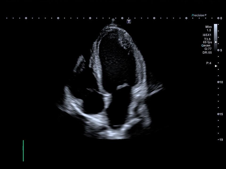
- What view or anatomy is shown?
- What probe would commonly be used?
- What is this exam used to evaluate?
Reveal Answer
Small footprint helps image between the ribs for cardiac views.
What Are You Looking At?
Before You Reveal
Study the image and answer these questions:
- What body area is being evaluated?
- What probe may be used?
- What clinical question can this help answer?
Reveal Answer
Linear probes provide high-resolution imaging of shallow structures.
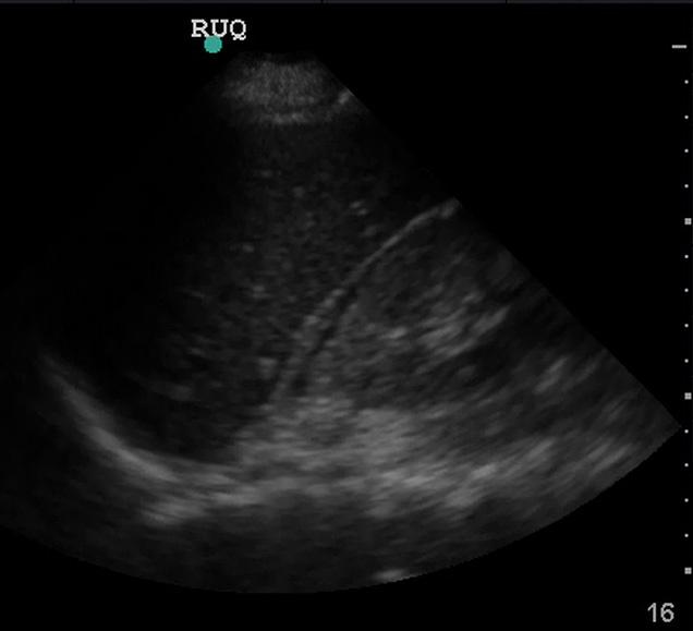
What Are You Looking At?
Before You Reveal
Study the image and answer these questions:
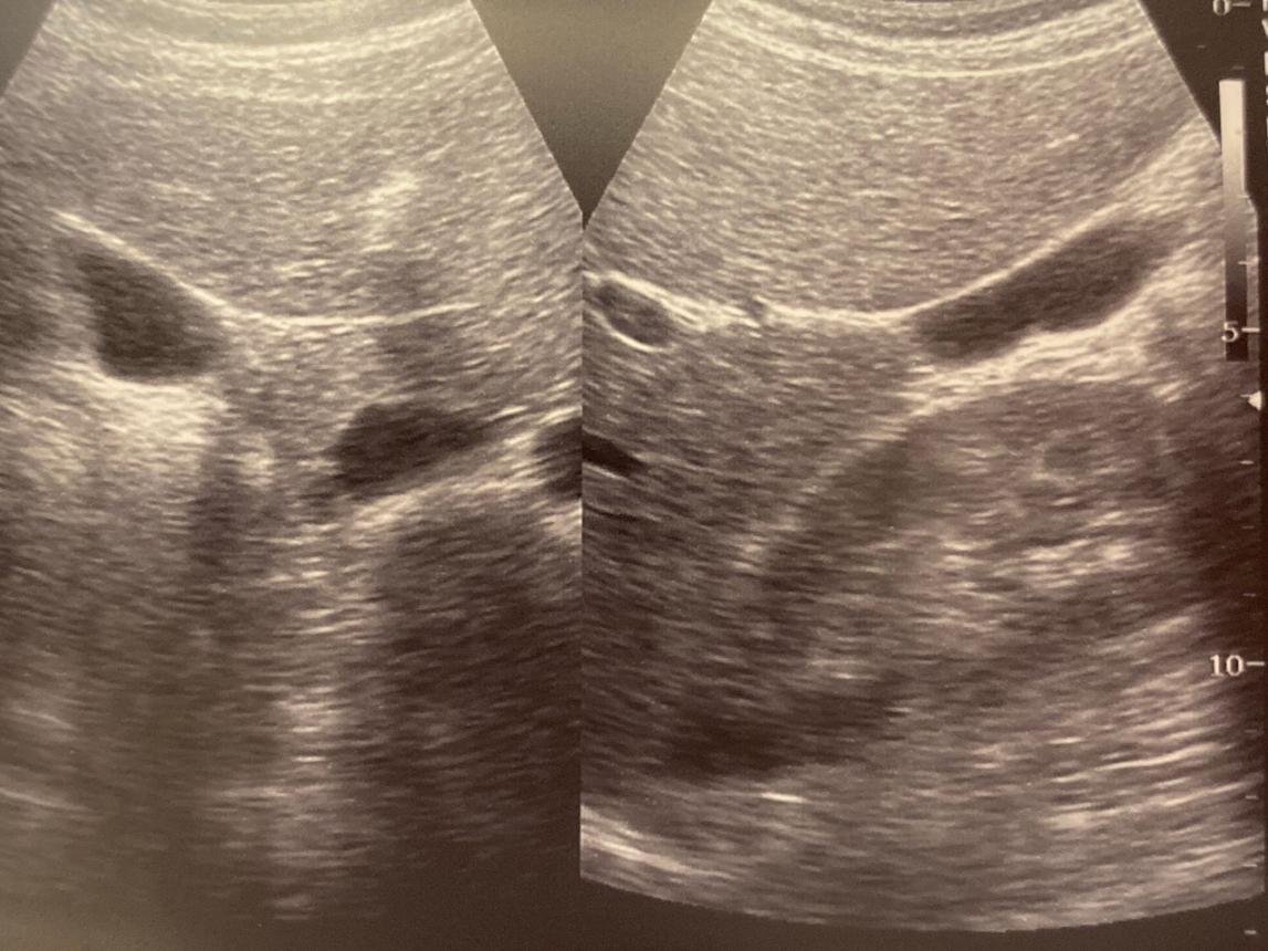
- What two structures are visible?
- What space is being evaluated?
- What probe is commonly used?
Reveal Answer
Curvilinear probes are commonly used for deeper abdominal imaging.
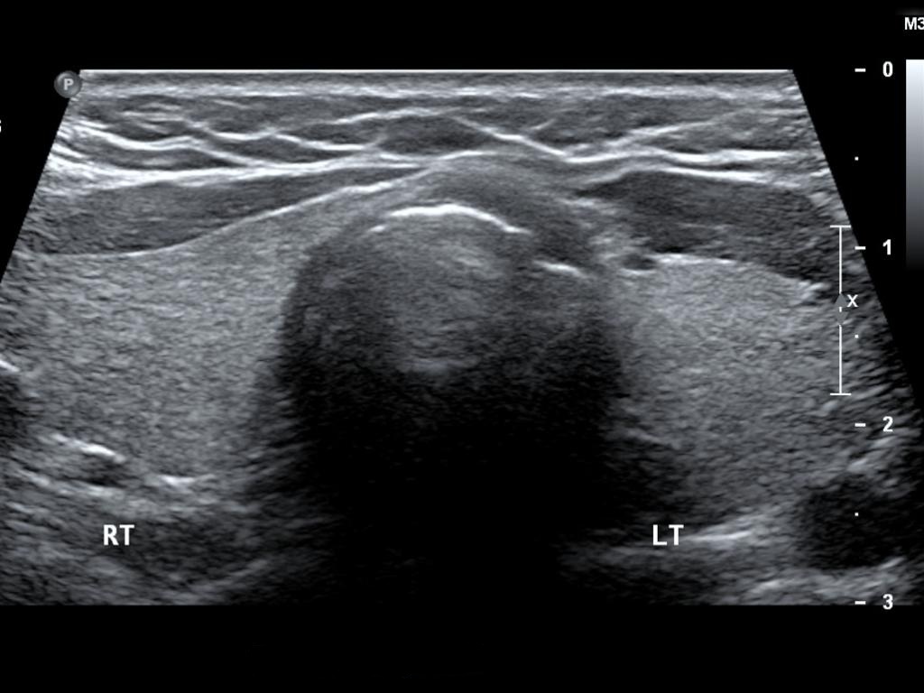
What Are You Looking At?
Before You Reveal
Study the image and answer these questions:
- What structure is shown?
- What probe is typically used?
- Why is this image usually high resolution?
Reveal Answer
Linear probes are ideal for thyroid and other shallow structures.
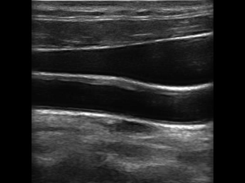
What Are You Looking At?
Before You Reveal
Study the image and answer these questions:
- Which structure is the vein?
- How can you tell the vein from the artery?
- What probe is commonly used?
Reveal Answer
Linear probes are commonly used for vascular access because the anatomy is superficial.
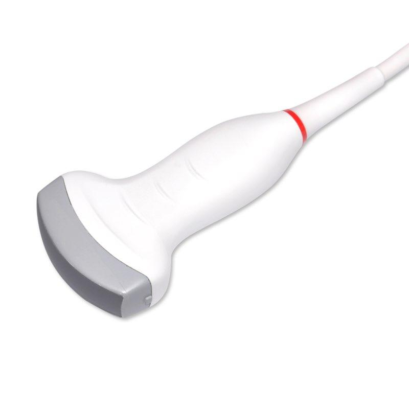
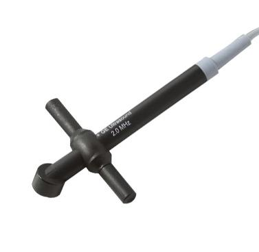
Which Probe Does Not Create an Image?
Before You Reveal
Study the probe and answer these questions:
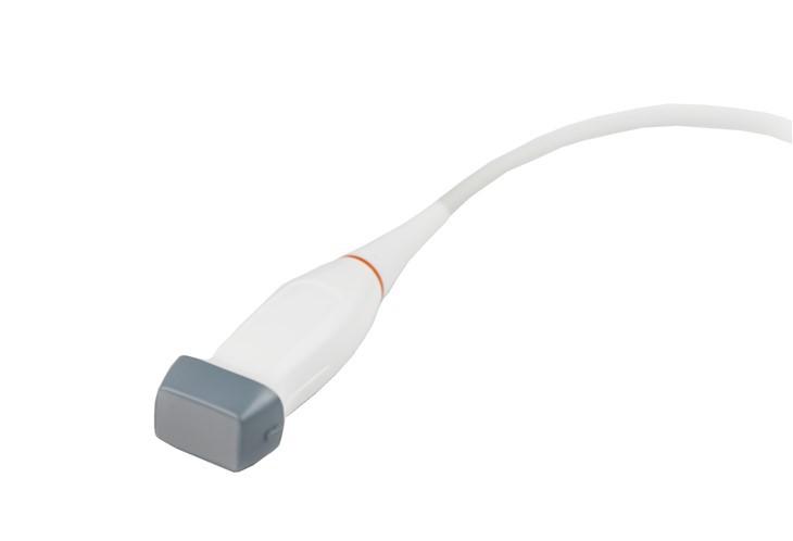
- What type of probe is this?
- Does it create a picture?
- What is it used to measure?
Reveal Answer
This probe does not create an image. It only detects blood flow velocity using sound.
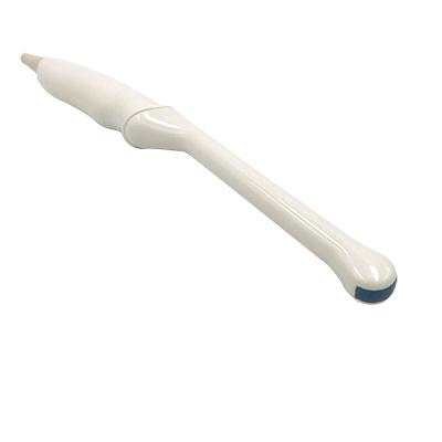
Which Probe Is Used Internally?
Before You Reveal
Study the probe and answer these questions:
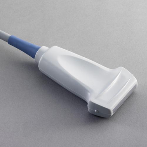
- What type of probe is this?
- Why does it provide high-resolution images?
- What exams commonly use this type of probe?
Reveal Answer
Used for internal imaging when close proximity to the anatomy gives better resolution.
Probe Notch and Image Orientation
The probe notch, groove, light, or marker is one of the most important orientation clues in ultrasound. It tells the operator which side of the probe corresponds to the screen indicator. If the image looks reversed or confusing, the first thing to check is often the probe notch.
Elite Ultrasound Quiz: Planes, Probes, Notches, TGC and Image Quality
This is a true thinking quiz. Pick an answer first, then check your answer. Teaching appears only after you answer so the quiz does not give itself away.
Question 1: Scanning Plane Challenge
You are scanning the internal jugular vein and carotid artery in short axis. The vessels appear mostly round on the screen. Which imaging plane are you most likely using?
Correct Answer: B. Transverse / short axis
Teaching: Short axis cuts across the vessel, so the IJ and carotid usually look round or oval.
What to notice first: Round vessels usually mean transverse or short-axis view.
Why A is wrong: Longitudinal views follow the vessel length, so the vessel appears tube-like instead of round.
Question 2: Orientation Challenge
You are scanning the IJ in transverse view. The probe notch is pointed toward the patient’s right. Which side of the screen usually represents the patient’s right side?
Correct Answer: A. Left side of the screen
Teaching: The notch corresponds to the screen indicator. If the notch points to patient right, patient right appears on the indicator side.
What to notice first: Always check the notch before deciding left vs right.
Why D is wrong: The notch is specifically there to give the image orientation.
Question 3: Probe Rotation Challenge
You rotate the probe 90 degrees and now the vessel appears as a long dark tube instead of a circle. What view are you most likely seeing?
Correct Answer: B. Longitudinal / long axis
Teaching: Long axis follows the length of the structure, so a vessel looks like a long tube.
What to notice first: Tube-like structure means long axis. Round structure means short axis.
Question 4: Probe Function Challenge
A small pencil-style probe on an echo machine produces sound and velocity information but no 2D picture. What probe is this?
Correct Answer: C. Continuous Wave Doppler probe
Teaching: CW Doppler measures blood flow velocity using sound and Doppler signal. It does not produce a 2D anatomy image.
What to notice first: Flow only, no image means CW Doppler.
Question 5: Probe Selection Challenge
You need high-resolution imaging of the thyroid gland. Which probe is the best starting choice?
Correct Answer: A. Linear probe
Teaching: The thyroid is superficial. A high-frequency linear probe gives better detail for shallow structures.
What to notice first: Superficial anatomy usually means linear probe.
Why B is wrong: Curvilinear probes are better for deeper abdomen, not shallow high-detail neck structures.
Question 6: Abdominal Imaging Challenge
You are trying to evaluate the liver and right kidney area for possible free fluid. Which probe is commonly used?
Correct Answer: A. Curvilinear probe
Teaching: RUQ FAST views often require deeper penetration to see abdominal anatomy.
What to notice first: Deep abdomen usually means curvilinear.
Question 7: Image Brightness Challenge
The top of the ultrasound image is bright, but the deeper part of the image is too dark. Which control is most directly designed to adjust brightness at different depths?
Correct Answer: A. TGC sliders
Teaching: Time gain compensation changes brightness by depth, helping balance near field and far field brightness.
What to notice first: Uneven brightness from top to bottom often points to TGC adjustment.
Question 8: Image Focus Challenge
You are imaging a target structure in the middle of the screen. Where should the focal zone usually be placed?
Correct Answer: A. At or just below the structure of interest
Teaching: The focal zone improves detail around a chosen depth, so place it near the anatomy you care about.
What to notice first: If the target is blurry, check focus placement.
Question 9: Image Quality Challenge
The image is weak and patchy because there is air between the probe and skin. What is the best first correction?
Correct Answer: A. Add gel and improve probe contact
Teaching: Air blocks ultrasound transmission. Gel removes air and improves coupling.
What to notice first: Dropout or patchy image can be a coupling problem.
Why D is wrong: Air is the enemy of ultrasound transmission at the probe-skin interface.
Question 10: Depth Challenge
The thyroid and IJ are usually located close to the probe. Which image region are they typically in?
Correct Answer: A. Near field
Teaching: The near field is the shallow region closest to the probe. Superficial structures appear there.
What to notice first: Shallow anatomy sits near the top of the image.
Question 11: Internal Probe Challenge
A long internal probe is used to get closer to pelvic anatomy and improve detail. What probe type is this?
Correct Answer: A. Endocavity probe
Teaching: Endocavity probes are used internally, placing the transducer closer to the anatomy for better resolution.
What to notice first: Long internal probe shape usually points to endocavity use.
Question 12: Vessel Compression Challenge
While scanning the IJ, you press too hard and the vein disappears. What likely happened?
Correct Answer: A. The vein compressed
Teaching: Veins are more compressible than arteries. Too much pressure can collapse the vein and make it hard to see.
What to notice first: If a vessel disappears with pressure, think vein compression.
Ultrasound Probe Quick Guide
Probe selection depends on the depth of the structure, the size of the imaging window, and the level of detail needed.
Phased Array
Best for cardiac imaging. The small footprint helps image between ribs.
Curvilinear
Best for deeper abdominal imaging such as RUQ, abdomen, and FAST exam views.
Linear
Best for superficial structures such as thyroid, vessels, IJ access, and vascular imaging.
Continuous Wave Doppler
Does not create a picture. It detects blood flow velocity using Doppler sound and is commonly used in echo.
Endocavity
Used for internal imaging such as pelvic, early pregnancy, and prostate evaluation where close detail is needed.
Ultrasound Image Optimization Basics
Image quality is not only about the probe. The operator also adjusts depth, gain, TGC, focal zone, and coupling to improve the image. These basics help learners understand why the same anatomy can look clear in one image and poor in another.
Gel and Proper Coupling
Ultrasound needs good contact between the probe and the skin. Gel removes air between the probe face and the patient. Without gel or proper coupling, the sound beam does not transmit well and the image can become weak, noisy, or incomplete.
Beginner Ultrasound Tips
Keep Practicing
Build confidence by connecting anatomy, probe selection, and clinical purpose. Try another MedSkillBuilder practice tool below.